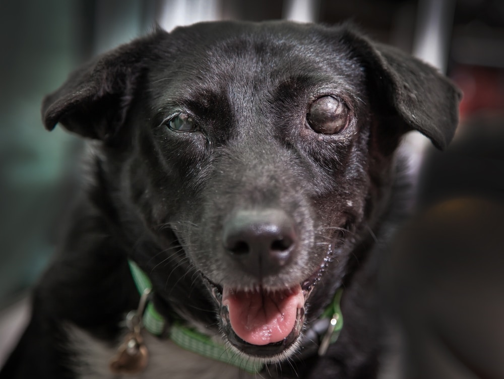
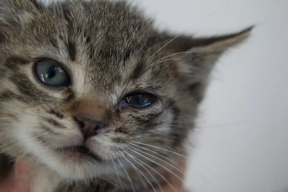
Many conditions, such as injury, infection, immune-mediated disorders, conformation abnormalities, and complications from other health issues, that can affect your pet’s eyes can potentially jeopardize their vision. Our Pets & Friends Animal Hospital team explains what eye issues commonly affect pets.
Corneal ulcers in pets
A corneal ulcer is a defect or abrasion in the corneal surface that can be caused by corneal trauma, foreign bodies, chronic corneal irritation, and chemical exposure. Most corneal ulcers involve only the superficial corneal tissue and heal quickly, but if the ulcer is deep or becomes infected, complications, such as eye rupture or blindness, can occur.
Corneal ulcer signs include excessive tearing, inability to open the eye, and eyelid swelling. The corneal surface may also appear cloudy and the sclera (i.e., white eye portion) may be red or blood-shot. A special stain (i.e., fluorescein) helps veterinarians detect corneal lesions, because the stain adheres only to the damaged area and highlights the lesion in a bright green color. Treatment typically includes topical eye medications, such as antibiotics and atropine, and systemic pain medications, because the ulcers are typically painful. In complicated cases, surgery to remove damaged tissue and repair the cornea may be necessary.
Keratoconjunctivitis sicca in pets
Keratoconjunctivitis sicca (KCS), commonly referred to as dry eye, is the result of inadequate eye lubrication because of the tear film’s insufficient aqueous portion. The most common cause is an immune-mediated disorder that damages the tear producing glands. Other causes include systemic infection, hypothyroidism, inner ear infection complications, and certain medications. Specific dog breeds, including cocker spaniels, bloodhounds, miniature schnauzers, pugs, and shih tzus, are at higher KCS risk.
KCS causes significant, chronic eye irritation with signs that include eye redness, squinting, and excessive blinking, and often a thick, yellowish mucoid discharge. Secondary corneal ulcers can also occur.
KCS is diagnosed using the Schirmer tear test, which measures tear production. Treatment involves stimulating tear production and replacing the tear film. Topical eye medications several times per day are usually required, and cleaning your pet’s eyes frequently with a warm, wet washcloth can help. KCS typically requires lifelong treatment.
Cherry eye in pets
A fibrous attachment typically anchors the third eyelid gland to the lower eye rim. In certain breeds, such as cocker spaniels, Boston terriers, beagles, pugs, and Persian cats, the attachment is weak, and the gland prolapses. This condition is called a cherry eye, because the prolapsed gland appears as a red, swollen mass on the lower eyelid near the muzzle.
The third eyelid gland produces about 50% of the tear film’s aqueous portion, so replacement is necessary to prevent KCS. In mild cases anti-inflammatories can correct the problem, but surgery is tyically necessary. If medical therapy isn’t working, surgery to replace the third eyelid gland should be performed as soon as possible to prevent permanent damage.
Entropion in pets
Entropion is a developmental or anatomic problem usually seen in young, rapidly growing dogs, such as bloodhounds, bull mastiffs, Chesapeake Bay retrievers, shar pei, and doberman pinschers. The eyelid rolls inward, the eyelid hair rubs the corneal surface, and the chronic irritation can lead to corneal inflammation and ulceration.
Affected pets typically have excessive tear production and hold their eyes closed, and some have a mucoid discharge. Treatment typically involves surgical correction to reverse the eyelid’s inward roll, although surgery is not usually performed until the pet is an adult to prevent over or under entropion correction. A veterinarian can place temporary sutures to roll the eyelids outward until the pet reaches the appropriate age. In some cases, the entropion resolves on its own as the pet grows.
Cataracts in pets
Cataracts are opacities that develop inside the translucent disc-like lens. Small cataracts may not cause a problem, but large cataracts can lead to blindness. Causes include chronic eye inflammation, genetics, age-related changes, eye trauma, and in dogs, diabetes. About 80% of diabetic dogs will eventually develop cataracts.
Cataracts’ typical signs include a cloudy or hazy appearance to one or both eyes, bumping into furniture, and hesitancy to move in unfamiliar settings. Fortunately, the cataracts themselves are not painful. Our veterinary team diagnoses cataracts by evaluating the eye interior, including the lens, with an ophthalmoscope. No medication can remove cataracts although ocular or systemic medications may delay progression in some cases. Surgery is the only way to remove cataracts.
Glaucoma in pets

Glaucoma, which is elevated intraocular pressure (IOP) caused by inadequate aqueous fluid drainage, can be primary, caused by inherited anatomical abnormalities in the drainage angle, or secondary, caused by issues such as chronic interior eye inflammation, lens dislocation, tumors, intraocular bleeding, and lens damage. Without relief, high IOP can damage the retina and optic nerve, potentially leading to blindness.
Initially, affected pets may show no signs, but as the IOP increases, signs may include eye rubbing, watery eye discharge, eye bulging, and a cloudy or bluish cornea. Our veterinary team diagnoses glaucoma using a tonometer applied appropriately to your pet’s cornea to measure IOP. Glaucoma treatment usually involves topical eye medications to reduce the IOP and analgesics to control pain. Surgical techniques may also be recommended in severe cases.
If your pet has eye discomfort, contact our Pets & Friends Animal Hospital team, so we can perform a thorough ophthalmic examination and devise a treatment plan to address the problem.

